(860) 430-9690
59 Sycamore St #301,Fax: (860) 430-9693
Glastonbury, CT 06033

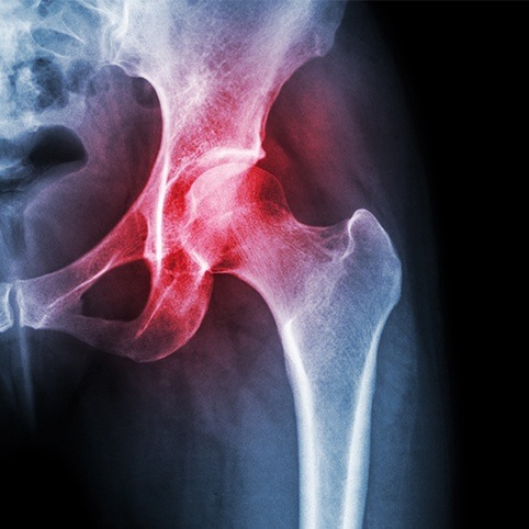
Femoroacetabular Impingement, or "FAI" as it’s known, is a condition where the ball of the femur (the hip bone) bangs up against, or impinges against, the lip of the socket (the acetabulum). Despite all that verbiage, it’s easy to understand why this condition would be painful! At Valley Sports Physicians & Orthopedic Medicine, we can identify the cause of a patient’s FAI using diagnostic ultrasound, and then provide treatments designed to reduce pain and improve mobility. We offer solutions for treating femoraacetabular (hip) impingement in Glastonbury, CT at Valley Sports Physicians & Orthopedic Medicine.

There are two main conventional causes of FAI:
A Cam Lesion occurs if the ball is not shaped normally, and the abnormal part of the ball can jam against the lip of the socket when the hip is bent.
A Pincer Lesion can develop if the front rim of the socket sticks out too far and it digs into the ball or neck of the femur during normal hip flexion movement.
But the bigger question is why did either of these lesions develop in the first place? The answer, and one which few physicians and surgeons discuss or are even aware of, is underlying instability of the hip. (Maybe because there's not much that conventional medicine can do to treat instability. But you'll see below that this isn't the case!) When the ligaments that support and stabilize and the joint become loose or overstretched, whether as a result of a specific hip injury or because of repetitive stress over time, it can allow the ball to slosh around in the socket. The excess movement can put more stress on the joint, resulting in bony overgrowth and giving rise to the cam and pincer lesions. (Think of attempting to drive a wooden stake into the ground with a hammer. Eventually, the force of the hammer blows causes the head of the stake to start to mushroom over and form "spurs" around the edges.)
To be fair, in rare cases cam and pincer lesions develop because there's a problem with both the ball and the socket:

By far the most common symptoms of FAI are:
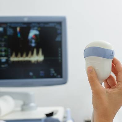
The patient history and physical exam usually give a strong clue to the diagnosis of impingement. But x-rays are required to confirm the presence of a cam and/or pincer lesion. One special x-ray view that most physicians don't order, but that can be particularly helpful in identifying FAI, and a Cam lesion in particular, is the Dunn View. However, studies have shown that people can have x-ray evidence of FAI, including Cam and/or Pincer lesions, and yet not have any symptoms of FAI! So, it's important to treat the patient and not the x-rays! In our office, we'll often perform a diagnostic injection to see if the impingement is actually causing the pain. Under ultrasound guidance, we inject the hip joint with some anesthetic. If the pain goes away then it's likely that the impingement is the cause.
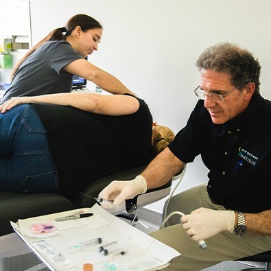
If the symptoms of FAI are mild, and the x-rays show relatively minimal changes, physical therapy is the first approach to treatment. But it's important to work with a therapist who can also identify and address any underlying instability. If the symptoms are more severe, and x-rays show large Cam and/or Pincer lesions, then arthroscopic surgery usually is needed to shave down the lesions and free up the movement. No physical therapy or injection treatment can get rid of the bony lesions. Underlying joint instability more often needs to be treated with Regenerative Medicine injection treatments such as Prolotherapy or PRP.* These treatments are designed to tighten up the loose ligaments and help stabilize the joint. Our physicians are highly skilled in Regenerative Medicine treatments and have been performing them longer than most in the country; few have our level of skill and experience.
(*NOTE: All Regenerative Medicine treatments are performed by our affiliate practice, the New England Stem Cell Institute. Click HERE for a free e-book on Regenerative Medicine.)